

The Mona Lisa and Sistine Chapel Keep “Cadavers” Alive
- Alessandro Berni
- Aug 16, 2018
- 7 min read
Artists create art because of the core reason that they love doing it. It is not merely for the viewer. First it is the process of creation that happens in an artist’s studio. It is a process of creation between the artist and his artwork. A place where the artist feels happy, the art relaxes mind, body and soul, something similar to meditation. Artists are the ones who are full of emotions, feelings, liveliness, happiness and joy. And that is what motivates them to create art. These words may sound a bit too spiritual but clearly relates to what Van Gogh had said about art "I put my heart and soul into my work, and have lost my mind in the process". A strong understanding of drawing and painting lay basic foundation in an artist’s life. No matter how scary it might seem, artists have gone to a great extent to study when it has come to drawings. This desire to perfect their drawing abilities has even gone to the level that they have studied human drawings from cadavers and they still do.
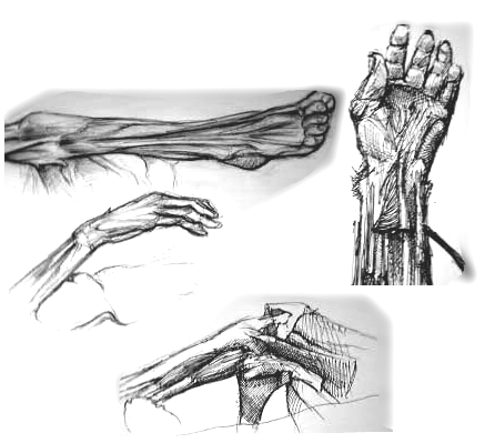
How many artists on an average among the entire world's population of artists, would have a fearless heart to enter into a science lab to study the dissecting cadavers? If given a thought it would be repulsive for most of them. An artist might not find it a beautiful thing to draw or paint. The sight and smell of the body, the sounds of cutting and sawing, and the feel of human flesh have effects both empathic and repulsive. But still there are artists and art students who do drawings from cadavers. This might seem a new concept of drawing but this trend has been alive since the 15th century, the Renaissance time period. Some examples from the present days have proven that the process of drawing from cadavers still exists.

The Art Students League of New York which has been instrumental in shaping America’s legacy in the fine arts as recently as this July conducted a workshop to study from cadavers. This course presented the study of anatomy as a convergence between anatomical and structural drawing. It motivated students of representational art and provided unparalleled opportunities for developing detailed anatomical knowledge through their work in Cornell College of Medicine’s anatomy lab.
The Drexel University College of Medicine and the Fleisher Art Memorial jointly offer “Advanced Artistic Anatomy,” a course consisting of 13 full days, which gives advanced art students the opportunity to directly study the cadavers dissected by medical students. This unique course focuses on the practical application of anatomical knowledge to drawing, painting and sculpture. It combines demonstrations and drawing in the anatomy lab with life drawing, where anatomical understanding is used to improve the student’s considerations of form and function, informing their approach to create artworks.
An art student shares his reaction after his drawing class from cadavers at a school in Florida “Here are some drawings I did in college of cadavers at a school in Florida. Wish I drew more but I was overwhelmed by the smell and left after 1 hour of sketching. I should have mentioned I did these almost 4 years ago. I think it's some of my best anatomy work from that year, my freshman year. I remember this one cross section of some guys head, that would have been a cool drawing but the smell and reality of what I was drawing was, again, too much for me. I would draw at least 20 sketches if I were to get the chance at cadavers again.”
Students in the art department at Eastern Illinois University had the opportunity to view and learn about different muscles from the inside of cadavers because of collaboration with biological sciences. Art professor Jenny Chi noted that her Advanced Level Life Drawing class had been learning and drawing from real-life models and learning about how the body was made. Chi said for the assignment, her students would have to view the cadavers and draw a section of the body. “I would love for my students to be able to do it all the time but it is not easy to access cadavers,” she said. Chi took some students last week to view the cadavers and to hear Thomas talk about them, but she did not know the response was going to be so positive. “We decided to do it again tonight, and this time students would go in with their sketchbooks and do an assignment,” Chi said. Chi expressed that although this is the first time art students have gotten to draw cadavers and she hopes this assignment can become part of the requirements for the class. Gary Bulla, chair of the biological sciences department, said this is the first time the two have collaborated. The cadavers can be challenging to get because the state provides a scarce amount of them, but the department was able to purchase two cadavers over the summer, Bulla said. Joshua Corry, a senior 2D studio art major, said that it is interesting to learn how the body works. “It is nice to see something that is underneath the skin and how it works. It is also nice to see what in underneath of what you are really drawing,” Corry said. Margaret Kilbane, a graduate student studying art, said that it was a cool experience to be able to see the internal organs because she is used to just seeing the external organs. Chi said for the assignment, her students would have to view the cadavers and draw a section of the body.
North Park University’s Cadavers Lab is another place where professors have used cadavers to teach drawing. It’s just another day in North Park University’s Cadaver Lab, When students gather and Visiting Assistant Professor of Biology Lindsey Alexander unzips a large body bag, carefully peeling back a sheet to reveal an arm. Gently parting the skin, Dr. Alexander points to various muscles as the anatomy students try to identify them. “Bicep?” says one student,“Flexors?” guesses another. At the center of the lab are two cadavers, or donors, as they are known here as a sign of respect. One is a 75-year-old man who died of congestive heart failure, the other a woman in her 60s whose cause of death is unknown. "What’s even more valuable, says Dr. Nelson, is the camaraderie that forms among the students as they work around a body. The cadaver lab isn’t just for health sciences students. Nelson noted that art professors have used the cadavers to teach drawing. "The students might study Michelangelo. Then they come here and see the real thing, and they might end up drawing it a little different than before,” Nelson added. Nelson also has invited students from nearby high schools such as Von Steuben and Northside College Prep into the lab. “We’re not the only university to have a cadaver lab,” says Nelson, “But the access we allow is not typical.” In addition, North Park students have more opportunities to actually dissect bodies, as opposed to schools where donor bodies arrive already cut open. The bodies often last for several years because of the special care Dr. Nelson takes to preserve them, covering them with dampened microfiber cloths after each session.
These students are lucky to not have to dissect the bodies themselves, where as artists during the Renaissance assisted in dissection while studying the cadavers.
Some artists even forged partnerships with specific physicians (Titian and Andreas Vesalius are perhaps the best- known example), in which the physicians would allow the artists to assist in dissections (highly restricted at the time) in exchange for anatomical drawings and illustrations. The Florentine sculptor Baccio Bandinelli, who appears to have run an academy for the teaching young artists has said, “I will show you that I know how to dissect the brain, and also living men, as I have dissected dead ones to learn my art”. Circumstantial evidence suggests that a number of other artists also attempted direct dissections.
Giorgio Vasari’s Lives of the Artists states that the great Florentine sculptor, painter, and printmaker Antonio Pollaiuolo who was the “first master to skin many human bodies in order to investigate the muscles and understand the nude in a more modern way.” Giving credence to Vasari’s claim, Pollaiuolo’s highly influential engraving of the Battle of Naked Men displays the figures of the nude warriors with nearly flayed musculature, seen in fierce action poses and from various angles.

Leonardo da Vinci and Michelangelo are also known to have undertaken detailed anatomical dissections at various points in their long careers. Leonardo da Vinci, who is without doubt the most significant artist-anatomist of all time, first undertook a series of detailed studies of the human skull in 1489, borrowing from the architect’s rigorous technique of representing three- dimensional forms in plan, section, elevation, and perspective view. He thereby invented a new vocabulary for the history of scientific illustration. Leonardo produced his most precisely drawn dissections of the human body in 1510–11, probably working under the direction of the young professor of anatomy, Marcantonio della Torre, from the University of Pavia. However, his methods of illustrating the dissection of muscles in layers, as well as some of his “plan, section, and elevation” techniques, seem to have become widely disseminated, and were incorporated in the first comprehensively illustrated Renaissance treatise, Andreas Vesalius’ De humani corporis fabrica, published in Basel in 1543. The Renaissance may be best known for its artworks: Michelangelo’s Sistine Chapel and “David,” and Da Vinci’s “Mona Lisa” and “Vitruvian Man” have without a doubt shaped the course of art history. Italian Renaissance artists became anatomists by necessity, as they attempted to refine a more lifelike, sculptural portrayal of the human figure. Indeed, until about 1500– 1510, their investigations surpassed much of the knowledge of anatomy that was taught at the universities.
In turn, physicians contracted artists to draw illustrations for the high volume of texts coming out in the field of anatomy, made possible by Gutenberg’s invention of the printing press around 1440.

Reading this history of art would of course motivate artists and art students to enter into biology labs in order to study cadavers. This gives confidence to those who are intrigued to attain perfection in their anatomical drawings in order to shape up their artistic career. And why not when one has an easy access in all legal ways along with good assistance it becomes easier to perfect his/her drawing skills. Who knows, how many Leonardos and Michelangelos are born in the time to come, who might take Mona Lisa and Sistine Chapel to another level of perfection! Since this is a highly advanced and technologically equipped time period, where there is access to anything and everything. This is the best time period for an artist to fully depict and express oneself in whatever form he/she wishes to, which can be well quoted as “freedom of expression”!
Comments